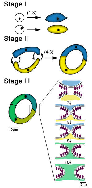
Figure 1. Three universal stages in the cell fusion pathway
(Stage I) Specification of the fusion fate
(1) Cells acquire specific fates.
(2) Selected cells execute their fates and in some cases proliferate.
(3) Cells undergo a differentiation program.
(Stage II) Cell attraction, attachment and recognition
(4) Cells destined to fuse find partners using migration mechanisms.
(5) Membrane micro-domains interact and form cell adhesion complexes.
(6) Cell signaling result in close recognition.
(Stage III) Execution of cell membrane and cytoplasmic fusion (a model)
(7) Fusion inducing proteins (FF proteins) localize to the cell membranes.
(8) Fusogens bend membranes and drive hemifusion of the outer membranes.
(9) Inner leaflets fuse forming pores, that expand and cytoplasm mix.
(10) The cytoplasmic contents (e.g. cytoskeleton) rearrange into a syncytium. (8)
Cell fusion is essential for sexual reproduction since fertilization requires the fusion of male and female gametes [1]. In addition, somatic cells fuse to sculpt organs such as in muscles, bones, placenta, eye lens and other organs throughout the animal kingdom [1]. However, little is known regarding the molecular mechanisms of cell fusion despite extensive attempts and a few notable exceptions [2]. Caenorhabditis elegans is a remarkable organism to study cell-cell fusion because more than one third of all the somatic cells and all the germ cells invariantly fuse during normal development in the skin, pharyngeal muscles, reproductive system, digestive system, excretory systems and nervous system. We have identified the first family of cellular fusion proteins (fusogens EFF-1 and AFF-1 in C. elegans) [3,4]. The current focus is determining the pattern and process by which cellular fusogens fuse eukaryotic cells during development.
EFF-1 (Epithelial Fusion Failure-1) and AFF-1 (Anchor cell Fusion Failure-1) are type I membrane proteins necessary and sufficient for cell fusion: eliminating fusion when absent and fusing cells when ectopically expressed on the membranes of the nematode C. elegans and heterologous cells [4,5]. We have shown that EFF-1 and AFF-1 must be expressed in both fusing cells, and thus that they act via novel homotypic interactions [6]. The fusion process shares a hemifusion intermediate with viral and intracellular membrane fusion processes (Fig. 1).
Homologs of EFF-1 and AFF-1 are present in over forty different nematode species [2], six arthropods, two ctenophores and one chordate. We hypothesize that diverse cell fusion events in nature will follow similar mechanisms as FF-mediated fusion. We will find the missing fusogens across kingdoms. Our evidence of the intermediates of the fusion process mediated by EFF-1 and AFF-1 in vitro and in vivo suggests that FF proteins fuse membranes by a mechanism analogous to viral fusogens or by a novel fusogenic paradigm [7]. Thus one present goal is to discover the mechanism of FF-mediated cell fusion.
References:
- Oren-Suissa, M. & Podbilewicz, B. Cell fusion during development. Trends Cell Biol 17, 537-546 (2007).
- Sapir, A., Avinoam, O., Podbilewicz, B. & Chernomordik, L. V. Viral and developmental cell fusion mechanisms: conservation and divergence. Dev Cell 14, 11-21 (2008).
- Mohler, W. A. et al. The type I membrane protein EFF-1 is essential for developmental cell fusion in C. elegans. Dev Cell 2, 355-362 (2002).
- Sapir, A. et al. AFF-1, a FOS-1-Regulated Fusogen, Mediates Fusion of the Anchor Cell in C. elegans. Dev Cell 12, 683-698 (2007).
- Podbilewicz, B. et al. The C. elegans developmental fusogen EFF-1 mediates homotypic fusion in heterologous cells and in vivo. Dev Cell 11, 471-481 (2006).
- Avinoam, O. et al. Conserved Eukaryotic Fusogens Can Fuse Viral Envelopes to Cells. Science 332, 589-592. (2011).
- Pérez-Vargas, J. et al. Structural basis of eukaryotic cell-cell fusion. Cell 157, 407-419 (2014).